Connect with us

The Conversation

How bird flu differs from seasonal flu − an infectious disease researcher explains

Published

on

theconversation.com – Hanna D. Paton, PhD Candidate in Immunology, University of Iowa – 2025-04-15 07:29:00

There is currently no bird flu vaccine for people.
Digicomphoto/ Science Photo Library via Getty Images

Hanna D. Paton, University of Iowa

The flu sickens millions of people in the U.S. every year, and the past year has been particularly tough. Although infections are trending downward, the Centers for Disease Control and Prevention has called the winter of 2024-2025 a “high severity” season with the highest hospitalization rate in 15 years.

Since early 2024, a different kind of flu called bird flu, formally known as avian influenza, has been spreading in birds as well as in cattle. The current bird flu outbreak has infected 70 Americans and caused two deaths as of April 8, 2025. Public health and infectious disease experts say the risk to people is currently low, but they have expressed concern that this strain of the bird flu virus may mutate to spread between people.

As a doctoral candidate in immunology, I study how pathogens that make us sick interact with our immune system. The viruses that cause seasonal flu and bird flu are distinct but still closely related. Understanding their similarities and differences can help people protect themselves and their loved ones.

What is influenza?

The flu has long been a threat to public health. The first recorded influenza pandemic occurred in 1518, but references to illnesses possibly caused by influenza stretch back as as early as 412 B.C., to a treatise called Of the Epidemics by the Greek physician Hippocrates.

Today, the World Health Organization estimates that the flu infects 1 billion people every year. Of these, 3 million to 5 million infections cause severe illness, and hundreds of thousands are fatal.

Influenza is part of a large family of viruses called orthomyxoviruses. This family contains several subtypes of influenza, referred to as A, B, C and D, which differ in their genetic makeup and in the types of infections they cause. Influenza A and B pose the largest threat to humans and can cause severe disease. Influenza C causes mild disease, and influenza D is not known to infect people. Since the turn of the 20th century, influenza A has caused four pandemics. Influenza B has never caused a pandemic.

An ad from 1918 for preventing influenza
A notice from Oct. 18, 1918, during the Spanish flu pandemic, about protecting yourself from infection.
Illustrated Current News/National Library of Medicine, CC BY

An influenza A strain called H1N1 caused the famous 1918 Spanish flu pandemic, which killed about 50 million people worldwide. A related H1N1 virus was responsible for the most recent influenza A pandemic in 2009, commonly referred to as the swine flu pandemic. In that case, scientists believe multiple different types of influenza A virus mixed their genetic information to produce a new and especially virulent strain of the virus that infected more than 60 million people in the U.S. from April 12, 2009, to April 10, 2010, and caused huge losses to the agriculture and travel industries.

Both swine and avian influenza are strains of influenza A. Just as swine flu strains tend to infect pigs, avian flu strains tend to infect birds. But the potential for influenza A viruses that typically infect animals to cause pandemics in humans like the swine flu pandemic is why experts are concerned about the current avian influenza outbreak.

Seasonal flu versus bird flu

Different strains of influenza A and influenza B emerge each year from about October to May as seasonal flu. The CDC collects and analyzes data from public health and clinical labs to determine which strains are circulating through the population and in what proportions. For example, recent data shows that H1N1 and H3N2, both influenza A viruses, were responsible for the vast majority of cases this season. Standard tests for influenza generally determine whether illness is caused by an A or B strain, but not which strain specifically.

Officials at the Food and Drug Administration use this information to make strain recommendations for the following season’s influenza vaccine. Although the meeting at which FDA advisers were to decide the makeup of the 2026 flu vaccine was unexpectedly canceled in late February, the FDA still released its strain recommendations to manufacturers.

The recommendations do not include H5N1, the influenza A strain that causes avian flu. The number of strains that can be added into seasonal influenza vaccines is limited. Because cases of people infected with H5N1 are minimal, population-level vaccination is not currently necessary. As such, seasonal flu vaccines are not designed to protect against avian influenza. No commercially available human vaccines currently exist for avian influenza viruses.

How do people get bird flu?

Although H5N1 mainly infects birds, it occasionally infects people, too. Human cases, first reported in 1997 in Hong Kong, have primarily occurred in poultry farm workers or others who have interacted closely with infected birds.

Initially identified in China in 1996, the first major outbreak of H5 family avian flu occurred in North America in 2014-2015. This 2014 outbreak was caused by the H5N8 strain, a close relative of H5N1. The first H5N1 outbreak in North America began in 2021 when infected birds carried the virus across the ocean. It then ripped through poultry farms across the continent.
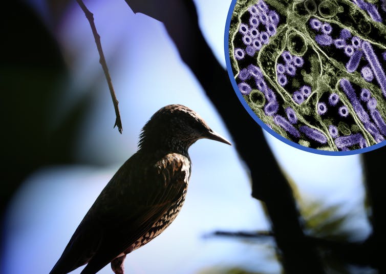
A bird and an image of H5N1 viral particles on a blue background
The H5N1 strain of influenza A generally infects birds but has infected people, too.
NIAID and CDC/flickr, CC BY

In March 2024, epidemiologists identified H5N1 infections in cows on dairy farms. This is the first time that bird flu was reported to infect cows. Then, on April 1, 2024, health officials in Texas reported the first case of a person catching bird flu from infected cattle. This was the first time transmission of bird flu between mammals was documented.

As of March 21, 2025, there have been 988 human cases of H5N1 worldwide since 1997, about half of which resulted in death. The current outbreak in the U.S. accounts for 70 of those infections and one death. Importantly, there have been no reports of H5N1 spreading directly from one person to another.

Since avian flu is an influenza A strain, it would show up as positive on a standard rapid flu test. However, there is no evidence so far that avian flu is significantly contributing to current influenza cases. Specific testing is required to confirm that a person has avian flu. This testing is not done unless there is reason to believe the person was exposed to sick birds or other sources of infection.

How might avian flu become more dangerous?

As viruses replicate within the cells of their host, their genetic information can get copied incorrectly. Some of these genetic mutations cause no immediate differences, while others alter some key viral characteristics.

Influenza viruses mutate in a special way called reassortment, which occurs when multiple strains infect the same cell and trade pieces of their genome with one another, potentially creating new, unique strains. This process prolongs the time the virus can inhabit a host before an infection is cleared. Even a slight change in a strain of influenza can result in the immune system’s inability to recognize the virus. As a result, this process forces our immune systems to build new defenses instead of using immunity from previous infections.

Reassortment can also change how harmful strains are to their host and can even enable a strain to infect a different species of host. For example, strains that typically infect pigs or birds may acquire the ability to infect people. Influenza A can infect many different types of animals, including cattle, birds, pigs and horses. This means there are many strains that can intermingle to create novel strains that people’s immune systems have not encountered before – and are therefore not primed to fight.

It is possible for this type of transformation to also occur in H5N1. The CDC monitors which strains of flu are circulating in order prepare for that possibility. Additionally, the U.S. Department of Agriculture has a surveillance system for monitoring potential threats for spillover from birds and other animals, although this capacity may be at risk due to staff cuts in the department.

These systems are critical to ensure that public health officials have the most up-to-date information on the threat that H5N1 poses to public health and can take action as early as possible when a threat is evident.The Conversation

Hanna D. Paton, PhD Candidate in Immunology, University of Iowa

This article is republished from The Conversation under a Creative Commons license. Read the original article.

Read More

The post How bird flu differs from seasonal flu − an infectious disease researcher explains appeared first on theconversation.com

The Conversation

The rise and fall – and rise again – of white-tailed deer

Published

on

theconversation.com – Elic Weitzel, Peter Buck Postdoctoral Research Fellow, Smithsonian Institution – 2025-05-29 07:48:00


White-tailed deer, once nearly extinct with only about 300,000 individuals a century ago, now number 30 to 35 million across North America. Historically, Indigenous hunting and land management, including controlled burns, supported stable deer populations for thousands of years. However, European colonization introduced commercial hunting for deer skins and meat, drastically increasing pressure and driving populations near extinction by the early 20th century. Conservation efforts like the 1900 Lacey Act and state protections ended commercial hunting, allowing deer numbers to recover through regulated hunting and reintroduction. This history highlights sustainable Indigenous practices versus later exploitative commercial pressures and modern conservation success.

White-tailed deer in North America are back to their precolonial population levels.
John Woodhouse Audubon/Heritage Images/Hulton Archive via Getty Images

Elic Weitzel, Smithsonian Institution

Given their abundance in American backyards, gardens and highway corridors these days, it may be surprising to learn that white-tailed deer were nearly extinct about a century ago. While they currently number somewhere in the range of 30 million to 35 million, at the turn of the 20th century, there were as few as 300,000 whitetails across the entire continent: just 1% of the current population.

This near-disappearance of deer was much discussed at the time. In 1854, Henry David Thoreau had written that no deer had been hunted near Concord, Massachusetts, for a generation. In his famous “Walden,” he reported that:

“One man still preserves the horns of the last deer that was killed in this vicinity, and another has told me the particulars of the hunt in which his uncle was engaged. The hunters were formerly a numerous and merry crew here.”

But what happened to white-tailed deer? What drove them nearly to extinction, and then what brought them back from the brink?

As a historical ecologist and environmental archaeologist, I have made it my job to answer these questions. Over the past decade, I’ve studied white-tailed deer bones from archaeological sites across the eastern United States, as well as historical records and ecological data, to help piece together the story of this species.

Precolonial rise of deer populations

White-tailed deer have been hunted from the earliest migrations of people into North America, over 15,000 years ago. The species was far from the most important food resource at that time, though.

Archaeological evidence suggests that white-tailed deer abundance only began to increase after the extinction of megafauna species like mammoths and mastodons opened up ecological niches for deer to fill. Deer bones become very common in archaeological sites from about 6,000 years ago onward, reflecting the economic and cultural importance of the species for Indigenous peoples.

three people crouched under deerskins with heads and antlers aim bows at several live deer
A 16th-century engraving of Indigenous Floridians hunting deer while disguised in deerskins.
Theodor de Bry/DEA Picture Library/De Agostini via Getty Images

Despite being so frequently hunted, deer populations do not seem to have appreciably declined due to Indigenous hunting prior to AD 1600. Unlike elk or sturgeon, whose numbers were reduced by Indigenous hunters and fishers, white-tailed deer seem to have been resilient to human predation. While archaeologists have found some evidence for human-caused declines in certain parts of North America, other cases are more ambiguous, and deer certainly remained abundant throughout the past several millennia.

Human use of fire could partly explain why white-tailed deer may have been resilient to hunting. Indigenous peoples across North America have long used controlled burning to promote ecosystem health, disturbing old vegetation to promote new growth. Deer love this sort of successional vegetation for food and cover, and thus thrive in previously burned habitats. Indigenous people may have therefore facilitated deer population growth, counteracting any harmful hunting pressure.

More research is needed, but even though some hunting pressure is evident, the general picture from the precolonial era is that deer seem to have been doing just fine for thousands of years. Ecologists estimate that there were roughly 30 million white-tailed deer in North America on the eve of European colonization – about the same number as today.

several people seated around rectangular hole showing dirt that's cut into grass
Elic Weitzel and volunteers excavate for deer bones at a 17th-century colonial site in Connecticut.
Scott Brady

Colonial-era fall of deer numbers

To better understand how deer populations changed in the colonial era, I recently analyzed deer bones from two archaeological sites in what is now Connecticut. My analysis suggests that hunting pressure on white-tailed deer increased almost as soon as European colonists arrived.

At one site dated to the 11th to 14th centuries – before European colonization – I found that only about 7% to 10% of the deer killed were juveniles.

Hunters generally don’t take juvenile deer if they’re frequently encountering adults, since adult deer tend to be larger, offering more meat and bigger hides. Additionally, hunting increases mortality on a deer herd but doesn’t directly affect fertility, so deer populations experiencing hunting pressure end up with juvenile-skewed age structures. For these reasons, this low percentage of juvenile deer prior to European colonization indicates minimal hunting pressure on local herds.

However, at a nearby site occupied during the 17th century – just after European colonization – between 22% and 31% of the deer hunted were juveniles, suggesting a substantial increase in hunting pressure.

one side of a deer's jaw bone with white teeth in place
Researchers can tell from the size and development of a deer’s bones its stage of life. Here is a fawn’s mandible with teeth.
Elic Weitzel

This elevated hunting pressure likely resulted from the transformation of deer into a commodity for the first time. Venison, antlers and deerskins may have long been exchanged within Indigenous trade networks, but things changed drastically in the 17th century. European colonists integrated North America into a trans-Atlantic mercantile capitalist economic system with no precedent in Indigenous society. This applied new pressures to the continent’s natural resources.

Deer – particularly their skins – were commodified and sold in markets in the colonies initially and, by the 18th century, in Europe as well. Deer were now being exploited by traders, merchants and manufacturers desiring profit, not simply hunters desiring meat or leather. It was the resulting hunting pressure that drove the species toward its extinction.

20th-century rebound of white-tailed deer

Thanks to the rise of the conservation movement in the late 19th and early 20th centuries, white-tailed deer survived their brush with extinction.

Concerned citizens and outdoorsmen feared for the fate of deer and other wildlife, and pushed for new legislative protections.

The Lacey Act of 1900, for example, banned interstate transport of poached game and – in combination with state-level protections – helped end commercial deer hunting by effectively de-commodifying the species. Aided by conservation-oriented hunting practices and reintroductions of deer from surviving populations to areas where they had been extirpated, white-tailed deer rebounded.

The story of white-tailed deer underscores an important fact: Humans are not inherently damaging to the environment. Hunting from the 17th through 19th centuries threatened the existence of white-tailed deer, but precolonial Indigenous hunting and environmental management appear to have been relatively sustainable, and modern regulatory governance in the 20th century forestalled and reversed their looming extinction.The Conversation

Elic Weitzel, Peter Buck Postdoctoral Research Fellow, Smithsonian Institution

This article is republished from The Conversation under a Creative Commons license. Read the original article.

Read More

The post The rise and fall – and rise again – of white-tailed deer appeared first on theconversation.com


Note: The following A.I. based commentary is not part of the original article, reproduced above, but is offered in the hopes that it will promote greater media literacy and critical thinking, by making any potential bias more visible to the reader –Staff Editor.

Political Bias Rating: Centrist

This content presents a balanced and fact-based historical and ecological account of white-tailed deer populations in North America. It avoids politically charged language or partisan framing, instead focusing on scientific findings, Indigenous land management practices, colonial impacts, and conservation efforts. The narrative recognizes both human influences and natural factors without attributing blame or praise in a manner aligned with any particular political ideology, reflecting a neutral and informative stance.

Continue Reading

The Conversation

There’s no evidence work requirements for Medicaid recipients will boost employment, but they are a key piece of Republican spending bill

Published

on

theconversation.com – Colin Gordon, Professor of History, University of Iowa – 2025-05-29 07:46:00


Republican senators in the U.S. Senate are debating their version of the budget and immigration bill passed by the House in May 2025. Some GOP senators want to reduce the $3.8 trillion deficit increase, while others oppose cuts to Medicaid, which serves low-income and disabled Americans. The House bill mandates work requirements for many Medicaid recipients by 2026, potentially denying coverage to millions. Historically, work requirements, rooted in 19th-century beliefs, have failed to promote employment or reduce poverty, instead causing harm and reducing access to benefits like Medicaid and SNAP. Courts have often ruled against Medicaid work requirements, viewing them as inconsistent with the program’s purpose. The House bill’s expansion of these requirements could leave 4.8 million uninsured.

Work requirements for receiving government benefits have a long history.
FatCamera/E+ via Getty Images

Colin Gordon, University of Iowa

Republicans in the U.S. Senate are sparring over their version of the multitrillion-dollar budget and immigration bill the House of Representatives passed on May 22, 2025.

Some GOP senators are insisting on shrinking the budget deficit, which the House version would increase by about US$3.8 trillion over a decade.

Others are saying they oppose the House’s cost-cutting provisions for Medicaid, the government’s health insurance program for people who are low income or have disabilities.

Despite the calls from U.S. Sen. Josh Hawley of Missouri and a few other Republican senators to protect Medicaid, as a scholar of American social policy I’m expecting to see the Senate embrace the introduction of work requirements for many adults under 65 who get health insurance through the program.

The House version calls for the states, which administer Medicaid within their borders and help pay for the program, to adopt work requirements by the end of 2026. The effect of this policy, animated by the conviction that coverage is too generous and too easy to obtain, will be to deny Medicaid eligibility to millions of those currently covered – leaving them without access to basic health services, including preventive care and the management of ongoing conditions such as asthma or diabetes.

Ending welfare

The notion that people who get government benefits should prove that they deserve them, ideally through paid labor, is now centuries old. This conviction underlay the Victorian workhouses in 19th-century England that Charles Dickens critiqued through his novels.

U.S. Rep. Brett Guthrie, R-Ky., put it bluntly earlier this month: Medicaid is “subsidizing capable adults who choose not to work,” he said.

People holding signs at a protest in support of Medicaid
Demonstrators in Illinois hold signs in support of Medicaid in 2018.
Charles Edward Miller via Wikimedia Commons, CC BY-SA

This idea also animated the development of the American welfare state, from its origins in the 1930s organized around the goals of maintaining civil order and compelling paid labor. Enforcing work obligations ensured the ready availability of low-wage labor and supported the growing assumption that only paid labor could redeem the lives and aspirations of the poor.

“We started offering hope and opportunity along with the welfare check,” Wisconsin Gov. Tommy Thompson argued in the early 1990s, “and expecting certain responsibilities in return.”

This concept also was at the heart of the U.S. government’s bid to end “welfare as we know it.”

In 1996, the Democratic Clinton administration replaced Aid to Families with Dependent Children, or AFDC, a long-standing entitlement to cash assistance for low-income families, with Temporary Aid for Needy Families, known commonly as TANF. The TANF program, as its name indicates, was limited to short-term support, with the expectation that most people getting these benefits would soon gain long-term employment.

Since 1996, Republicans serving at the state and federal levels of government have pressed to extend this principle to other programs that help low-income people. They’ve insisted, as President Donald Trump put it halfway through his first term, that unconditional benefits have “delayed economic independence, perpetuated poverty, and weakened family bonds.”

Such claims are unsupported. There is no evidence to suggest that work requirements have ever galvanized independence or lifted low-income people out of poverty. Instead, they have punished low-income people by denying them the benefits or assistance they require.

Work requirements haven’t worked

Work requirements have consistently failed as a spur to employment. The transition from the AFDC to TANF required low-income families to meet work requirements, new administrative burdens and punitive sanctions.

The new work expectations, rolled out in 1997, were not accompanied by supporting policies, especially the child care subsidies that many low-income parents with young children require to hold a job. They were also at odds with the very low-paying and unstable jobs available to those transitioning from welfare.

Scholars found that TANF did less to lift families out of poverty than it did to shuffle its burden, helping the nearly poor at the expense of the very poor.

The program took an especially large toll on low-income Black women, as work requirements exposed recipients to long-standing patterns of racial and gender discrimination in private labor markets.

Restricting access to SNAP

Work requirements tied to other government programs have similar track records.

The Supplemental Nutrition Assistance Program, which helps millions of Americans buy groceries, adopted work requirements for able-bodied adults in 1996.

Researchers have found that SNAP’s work requirements have pared back eligibility without any measurable increase in labor force participation.

As happens with TANF, most people with SNAP benefits who have to comply with SNAP work requirements are already working to the degree their personal circumstances and local labor markets allow.

The requirements don’t encourage SNAP recipients to work more hours; they simply lead people to be overwhelmed by red tape and stop renewing their SNAP benefits.

Failing in Arkansas

The logic of work requirements collapses entirely when extended to Medicaid.

Red states have been pressing for years for waivers that would allow them to experiment with work requirements – especially for the abled-bodied, working-age adults who gained coverage under the Affordable Care Act’s Medicaid expansion.

The first Trump administration granted 13 such waivers for what it saw as “meritorious innovations,” building “on the human dignity that comes with training, employment and independence.”

YouTube video
The House passed the budget bill on May 22, 2025. It includes steep cuts to Medicaid and imposes work requirements for eligibility.

Arkansas got the furthest with adding work requirements to Medicaid at that time. The results were disappointing.

“We found no evidence that the policy succeeded in its stated goal of promoting work,” as one research team concluded, “and instead found substantial evidence of harm to health care coverage and access.”

The Biden administration slowed down the implementation of these waivers by directing the Centers for Medicare and Medicaid Services to suspend or stem any state programs that eroded coverage. Meanwhile, state courts consistently ruled against the use of Medicaid work requirements.

In Trump’s second term, Iowa, Arizona and at least a dozen other states have proposed “work requirement” waivers for federal approval.

Trying it again

The waiver process is meant to allow state experiments to further the statutory objectives of the Medicaid program, which is to furnish “medical assistance on behalf of families with dependent children and of aged, blind, or disabled individuals, whose income and resources are insufficient to meet the costs of necessary medical services.”

On these grounds, the courts have consistently held that state waivers imposing work requirements not only fail to promote Medicaid’s objectives but amount to an arbitrary and capricious effort to undermine those objectives.

“The text of the statute includes one primary purpose,” the D.C. Circuit ruled in 2020, “which is providing health care coverage without any restriction geared to healthy outcomes, financial independence or transition to commercial coverage.”

Changing Medicaid in all states

The House spending bill includes a work requirement that would require all able-bodied, childless adults under 65 to demonstrate that they had worked, volunteered or participated in job training for 80 hours in the month before enrollment.

It would also allow states to extend such work requirements to six months and apply the new requirements not just to Medicaid recipients but to people who get subsidized health insurance through an Affordable Care Act exchange.

If passed in some form by the Senate, the House spending bill would transform the landscape of Medicaid work requirements, pushing an estimated 4.8 million Americans into the ranks of the uninsured.The Conversation

Colin Gordon, Professor of History, University of Iowa

This article is republished from The Conversation under a Creative Commons license. Read the original article.

Read More

The post There’s no evidence work requirements for Medicaid recipients will boost employment, but they are a key piece of Republican spending bill appeared first on theconversation.com


Note: The following A.I. based commentary is not part of the original article, reproduced above, but is offered in the hopes that it will promote greater media literacy and critical thinking, by making any potential bias more visible to the reader –Staff Editor.

Political Bias Rating: Center-Left

This article presents a critical perspective on Republican-led budget and Medicaid work requirement proposals, emphasizing their negative impacts on low-income populations and Medicaid coverage. It references academic research and historical context to argue that work requirements have failed to achieve stated goals and instead restrict access to essential health services. While it reports GOP positions and quotes Republican officials, the framing and choice of evidence highlight skepticism about conservative policy claims and focus on social justice and health equity concerns. This indicates a center-left bias oriented toward protecting government social programs and criticizing austerity measures.

Continue Reading

The Conversation

Billy Joel has excess fluid in his brain – a neurologist explains what happens when this protective liquid gets out of balance

Published

on

theconversation.com – Danielle Wilhour, Assistant Professor of Neurology, University of Colorado Anschutz Medical Campus – 2025-05-28 07:53:00


Cerebrospinal fluid (CSF) is a clear liquid that cushions the brain and spinal cord, supplies nutrients, and removes waste. Disorders like normal pressure hydrocephalus, recently diagnosed in Billy Joel, involve excess CSF accumulating in brain cavities, causing pressure despite normal diagnostic readings. CSF is produced mainly by the choroid plexus and circulates through brain ventricles and spinal canal. Problems with CSF flow can cause conditions like CSF leaks, which cause positional headaches and other symptoms, and idiopathic intracranial hypertension, characterized by increased skull pressure often linked to obesity. Treatments range from rest and caffeine to surgical shunts, aiming to reduce pressure and protect brain function. Further research is needed to fully understand CSF disorders.

Billy Joel was diagnosed with normal pressure hydrocephalus.
Ethan Miller/Getty Images

Danielle Wilhour, University of Colorado Anschutz Medical Campus

Cerebrospinal fluid, or CSF, is a clear, colorless liquid that plays a crucial role in maintaining the health and function of your central nervous system. It cushions the brain and spinal cord, provides nutrients and removes waste products.

Despite its importance, problems related to CSF often go unnoticed until something goes wrong.

Recently, cerebrospinal fluid disorders drew public attention with the announcement that musician Billy Joel had been diagnosed with normal pressure hydrocephalus. In this condition, excess CSF accumulates in the brain’s cavities, enlarging them and putting pressure on surrounding brain tissue even though diagnostic readings appear normal. Because normal pressure hydrocephalus typically develops gradually and can mimic symptoms of other neurodegenerative diseases, such as Alzheimer’s or Parkinson’s disease, it is often misdiagnosed.

I am a neurologist and headache specialist. In my work treating patients with CSF pressure disorders, I have seen these conditions present in many different ways. Here’s what happens when your cerebrospinal fluid stops working.

What is cerebrospinal fluid?

CSF is made of water, proteins, sugars, ions and neurotransmitters. It is primarily produced by a network of cells called the choroid plexus, which is located in the brain’s ventricles, or cavities.

The choroid plexus produces approximately 500 milliliters (17 ounces) of CSF daily, but only about 150 milliliters (5 ounces) are present within the central nervous system at any given time due to constant absorption and replenishment in the brain. This fluid circulates through the ventricles of the brain, the central canal of the spinal cord and the subarachnoid space surrounding the brain and spinal cord.

Diagram of cross-section of brain depicting cerebrospinal fluid circulation
Cerebrospinal fluid circulates throughout the brain and spinal cord.
OpenStax, CC BY-SA

CSF has several critical functions. It protects the brain and spinal cord from injury by absorbing shocks. Suspending the brain in this fluid reduces its effective weight and prevents it from being crushed under its own mass. Additionally, CSF helps maintain a stable chemical environment in the central nervous system, facilitating the removal of metabolic waste and the distribution of nutrients and hormones.

If the production, circulation or absorption of cerebrospinal fluid is disrupted, it can lead to significant health issues. Two notable conditions are CSF leaks and idiopathic intracranial hypertension.

Cerebrospinal fluid leak

A CSF leak occurs when the fluid escapes through a tear or hole in the dura mater – the tough, outermost layer of the meninges that surrounds the brain and spinal cord.

The dura can be damaged from head injuries or punctured during surgical procedures involving the sinuses, brain or spine, such as lumbar puncture, epidurals, spinal anesthesia or myelogram. Spontaneous CSF leaks can also occur without any identifiable cause.

CSF leaks were originally thought to be relatively rare, with an estimated annual incidence of 5 per 100,000 people. However, with increased awareness and advances in imaging, health care providers are discovering more and more leaks. They tend to occur more frequently in middle-aged adults and are more common in women than men.

Risk factors for the condition include connective tissue disorders such as Ehlers-Danlos syndrome as well as postural orthostatic tachycardia syndrome.

YouTube video
An upright headache could be a sign of a CSF leak.

Unfortunately, it’s common for health care providers to misdiagnose a CSF leak as another condition, like migraine, sinus infections or allergies. What can make diagnosing a CSF leak challenging is its broad symptoms. Most people with a CSF leak have a positional headache that improves when lying down and worsens when standing. Pain is usually felt in the back of the head and may involve the neck and between the shoulder blades. In addition to headaches, patients may experience ringing in the ears, vision disturbances, memory problems, brain fog, dizziness and nausea.

Imaging may help guide diagnosis, including an MRI of your brain or entire spine, or a myelogram of the space surrounding your spinal cord. Features of a CSF leak that are visible in a scan include your brain sagging down in the base of your skull as well as a fluid collection outside of your dura. However, an estimated 19% of people with a CSF leak can have normal scans, so not seeing signs of a leak on imaging does not entirely rule it out.

Conservative treatment for a CSF leak involves rest, lying flat and increasing your fluid intake to give your spine time to heal the puncture. Increasing your caffeine consumption to an equivalent of three to four cups of coffee per day can also help by increasing CSF production through stimulating the choroid plexus. Caffeine also relieves pain by interacting with adenosine receptors, which are key players in the body’s pain perception mechanisms.

If a conservative approach is not successful, an epidural blood patch may be necessary. In this procedure, blood is drawn from your arm and injected into your spine. The injected blood can help form a covering over the hole and promote the healing process. Headache improvement can be fast, but if the patch does not work or the results are short-lived, additional testing may be needed to better locate the site of the leak. In rare cases, surgery may be recommended. Most patients with a CSF leak respond to some form of these treatments.

Idiopathic intracranial hypertension

Idiopathic intracranial hypertension is a disorder involving an excess of CSF that elevates pressure inside the skull and compresses the brain. The term “idiopathic” indicates that the cause of the raised pressure is unknown.

Most patients with idiopathic intracranial hypertension have a history of obesity or recent weight gain. Other risk factors include taking certain medications such as tetracycline, excessive vitamin A, tretinoin, steroids and growth hormone. Middle-aged obese women are 20 times more likely to be diagnosed with idiopathic intracranial hypertension than other patient groups. As obesity becomes more prevalent, so too does the incidence of this condition.

YouTube video
Idiopathic intracranial hypertension results from increased intracranial pressure.

Patients with idiopathic intracranial hypertension typically experience headaches and vision changes, tinnitus or eye pain. Papilledema, or swelling of the optic disc, is the hallmark finding on a fundoscopic examination of the back of the eye. Clinicians may also observe paralysis of the patient’s eye muscles.

Normal pressure hydrocephalus, Joel’s diagnosis, is a form of this condition that commonly results in difficulty walking, loss of bladder control and cognitive impairment, sometimes referred to as the “wet, wobbly and wacky” triad. Joel’s diagnosis has brought awareness to this underrecognized but potentially treatable disorder, which is often managed through surgically placing a shunt to divert excess fluid and relieve symptoms.

Brain imaging of patients suspected of having idiopathic intracranial hypertension is crucial to excluding other causes of elevated CSF pressure, such as brain tumors or blood clots in the brain. A lumbar puncture or spinal tap to measure the pressure and composition of CSF is also central to diagnosis.

Since high intracranial pressure can damage the optic nerve and lead to permanent vision loss, the primary goal of treatment is to decrease pressure and preserve the optic nerve. Treatment options include weight loss, dietary changes and medications to reduce CSF production. Surgical procedures can also reduce intracranial pressure.

Future directions and unknowns

Cerebrospinal fluid is indispensable for brain health. Despite advances in understanding diseases related to CSF, several aspects remain unclear.

The exact mechanisms that lead to conditions like CSF leaks and idiopathic intracranial hypertension are not fully understood, though there are many theories. Further research is vital to enhance diagnostic accuracy and effective treatments for CSF disorders.

This is an updated version of an article originally published on Aug. 14, 2024.The Conversation

Danielle Wilhour, Assistant Professor of Neurology, University of Colorado Anschutz Medical Campus

This article is republished from The Conversation under a Creative Commons license. Read the original article.

Read More

The post Billy Joel has excess fluid in his brain – a neurologist explains what happens when this protective liquid gets out of balance appeared first on theconversation.com


Note: The following A.I. based commentary is not part of the original article, reproduced above, but is offered in the hopes that it will promote greater media literacy and critical thinking, by making any potential bias more visible to the reader –Staff Editor.

Political Bias Rating: Centrist

This content is purely scientific and medical in nature, focusing on cerebrospinal fluid disorders and their clinical aspects. It presents factual information, medical terminology, and treatment options without any political commentary or ideological framing. The neutral tone and emphasis on evidence-based medicine indicate no discernible political bias.

Continue Reading

Trending