fbpx
Connect with us

The Conversation

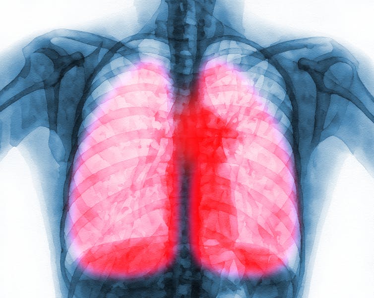
Long COVID inflicts deep scars on the lungs, but targeting specific immune cells could reverse damage − new research in mice

Published

on

theconversation.com – Harish Narasimhan, Ph.D. Candidate in Immunology, University of Virginia – 2024-09-04 10:05:22

Many respiratory viral infections can cause long-term symptoms.

sbk_20d pictures/Moment via Getty Images

Harish Narasimhan, University of Virginia

The long-term effects of respiratory viral infections such as are a major public health burden. Some estimates suggest over 65 million people around the world suffer from long COVID-19.

Advertisement

Efforts to better understand this , however, have been hampered by its ability to affect multiple organ systems, such as those involving the lungs, brain and heart. This is further complicated by the lack of animal models that can sufficiently mimic the disease.

Animal models, such as mice and rats, are a crucial tool that researchers use to study human diseases and develop treatment strategies. Although there are major differences between humans and animal models, the vast majority of our immune and organs function similarly. Such similarities in physiology have made significant discoveries, those related to COVID-19, possible.

I am an immunology researcher in the Sun Lab at the University of Virginia. We study the role the immune system plays in respiratory viral infections such as influenza and COVID-19. In our newly published research, we developed a new mouse model to study long COVID-19 and found that blocking certain overactive immune cells can restore lung function.

New models, new targets

Out team wanted to better understand the long-term effects of COVID-19 on the respiratory system. To do this, we worked to identify key features associated with lung scarring following COVID-19.

Advertisement

First, we examined lung samples from with long COVID-19. Although these patients were infected several months to years before the samples were taken, we found evidence of an overactive immune system in their lungs, particularly within that failed to fully repair themselves after infection.

Next, we aimed to create a mouse model for long COVID-19 by comparing the pathology of mice infected with four different types of respiratory viral infections. Surprisingly, we found that mice infected with influenza virus, rather than the COVID-19 mouse models scientists currently use, best replicated the physical features of severe chronic lung disease. The reasons why infections from different respiratory viruses affect the lungs in different ways are unclear. But preliminary evidence suggests it may be because each virus targets different types of cells “in humans and mice.”

Additionally, since long COVID-19 is about the left behind after infection, it seems less important what virus causes the problem in our animal model than that the damage is similar to what we want to address in human patients.

Long COVID-19 can be debilitating.

Using our new mouse model, we were able to identify the presence of an abnormal cluster of cells in mice lungs – made up of the same dysfunctional immune and epithelial, or structural, cells seen in the lungs of long-COVID-19 patients. Additionally, we found that the uncontrolled activity of these immune cells in the lungs impeded structural cells from repairing themselves. It also hindered them from restoring gas exchange, the of taking in oxygen and releasing carbon dioxide.

Advertisement

Importantly, when we blocked the activity of proteins associated with this overactive immune response, it reduced lung scarring and restored optimal lung function in mice.

Treating respiratory viral infections

Most approaches to addressing long COVID-19 rely on starting treatment early after infection. To the best of our knowledge, our study is the first to identify strategies to treat the respiratory symptoms of long COVID-19 after this chronic disease develops.

The we tested in our study have already been approved by the Food and Drug Administration to treat severe COVID-19 and other inflammatory conditions. We hope our findings can spur further research on using these drugs to treat long COVID-19.

Our work may have applications beyond long COVID-19. Growing evidence suggests that many respiratory viral infections, such as influenza, COVID-19 and respiratory syncytial virus, may result in chronic lung disease. Considering the four pandemics and even more respiratory viral epidemics that have occurred in the past 100 years, studying the cellular and molecular similarities between respiratory viral infections may be critical to how medical practitioners respond to future viral outbreaks.The Conversation

Harish Narasimhan, Ph.D. Candidate in Immunology, University of Virginia

Advertisement

This article is republished from The Conversation under a Creative Commons license. Read the original article.
Read More

The post Long COVID inflicts deep scars on the lungs, but targeting specific immune cells could reverse damage − new research in mice appeared first on theconversation.com

Advertisement

The Conversation

New FDA rule will ensure all women have more information after cancer screenings

Published

on

theconversation.com – Nancy Kressin, Emeritus Professor of Medicine, Boston – 2024-09-09 07:24:09

Breast density raises the risk of breast cancer and can also make it more difficult for breast cancer to be detected.
picture alliance/Getty Images

Nancy Kressin, Boston University; Christine M. Gunn, Dartmouth College; Priscilla J. Slanetz, Boston University, and Tracy A. Battaglia, Yale University

The Food and Drug Administration implemented a rule to go into effect on Sept. 10, 2024, requiring mammography facilities to notify women about their breast density. The goal is to ensure that women nationwide are informed about the risks of breast density, advised that other imaging tests might help find cancers and urged to talk with their about next steps based on their individual situation.

The FDA originally issued the rule on March 10, 2023, but extended the implementation date to give mammography facilities additional time to adhere.

Advertisement

The Conversation U.S. asked a team of experts in social science and patients’ health behaviors, health policy, radiology and primary care and health services research to explain the FDA’s new regulations about these health communications and what women should consider as they decide whether to pursue additional imaging tests, often called supplemental screening.

What is breast density and why does it matter?

Breast density is categorized into four categories: fatty, scattered tissue, heterogeneously dense or extremely dense.

Dense breasts are composed of more fibrous, connective tissue and glandular tissue – meaning glands that produce milk and tubes that carry it to the nipple – than fatty tissue. Because fibroglandular tissue and breast masses both look white on mammographic images, greater breast density makes it more difficult to detect cancer. Nearly half of all American women are categorized as dense breasts.

Having dense breasts also increases the risk of getting breast cancer, though the reason for this is unknown.

Advertisement

Because of this, decisions about breast cancer screening get more complicated. While evidence is clear that regular mammograms save lives, additional testing such as ultrasound, MRI or contrast-enhanced mammography may be warranted for women with dense breasts.

What does the new FDA rule say?

The FDA now requires specific language to ensure that all women receive the same “accurate, complete and understandable breast density information.” After a mammogram, women must be informed:

– Whether their breasts are dense or not dense

– That having dense breasts increases the risk of breast cancer

Advertisement

– That having dense breasts makes it harder to find breast cancer on mammograms

– That for those with dense breasts, additional imaging tests might help find cancer

They must also be advised to discuss their individual situation with their provider, to determine which, if any, additional screening might be indicated.

A female doctor holds a breast model up, explaining it to her patient, with a mammogram image in the background.
Conversations between patients and doctors are crucial for determining whether supplemental screening would be beneficial.
PonyWang/E+ via Getty Images

Why did the FDA issue the new rule?

Prior to the federal rule, 38 U.S. states required some form of breast density notification. But some states had no notification requirements, and among the others there were many inconsistencies that raised concerns by advocates, women with dense breasts whose advanced cancer had not been detected on a mammogram.

The FDA standardized the information women must receive. It is written at an eighth grade reading level and may address racial and literacy-level differences in women’s knowledge about breast density and reactions to written notifications.

Advertisement

For instance, our research team found disproportionately more confusion and anxiety among women of color, those with low literacy and women for whom English was not their first language. And some women with low literacy reported decreased future intentions to undergo mammographic screening.

What is the value of additional screening?

Standard mammograms use X-rays to produce two-dimensional images of the breast. A newer type of mammography imaging called tomosynthesis produces 3D images, which find more cancers among women with dense breasts. So, researchers and doctors generally agree that women with dense breasts should undergo tomosynthesis screening when available.

There is still limited scientific evidence to guide recommendations for supplemental breast screening beyond standard mammography or tomosynthesis for women with dense breast tissue. Data shows that supplemental screening with ultrasound, MRI or contrast-enhanced mammography may detect additional cancers, but there are no prospective studies confirming that such additional screening saves more lives.

So far, there is no data from randomized clinical trials showing that supplemental breast MRIs, the most often-recommended supplemental screening, reduce death from breast cancer.

Advertisement

However, more early stage – but not late-stage – cancers are found with MRIs, which may require less extensive surgery and less chemotherapy.

Various professional and experts interpret the available data about supplemental screening differently, arriving at different conclusions and recommendations. An important consideration is the woman’s individual level of risk, since emerging evidence suggests that women whose personal risk of developing breast cancer is high are most likely to benefit from supplemental screening.

Some organizations have concluded that current evidence is too limited to make a recommendation for supplemental screening, or they do not recommend routine use of supplemental screening for women based solely on breast density. Others recommend additional screening for women with extremely or heterogeneously dense breasts, even when their risk is at the intermediate level.

What should women consider about added screening?

Because personal risk of breast cancer is a crucial consideration in deciding whether to undergo supplemental screening, women should understand their own risk.

Advertisement

The American College of Radiology recommends that all women undergo risk assessment by age 25. Women and their providers can use risk calculators such as Tyrer-Cuzick, which is free and available online.

Women should also understand that breast density is only one of several risks for breast cancer, and some of the others can be modified. Engaging in regular physical activity, maintaining a healthy weight, limiting alcohol use and eating a healthy diet rich in vegetables can all decrease breast cancer risk.

Are there potential harms?

Amid the debate about the benefits of supplemental breast screening, there is less discussion about its possible harms. Most common are false alarms: results that suggest a finding of cancer that require follow-up testing. Less commonly, a biopsy is needed, which may to short-term fear and anxiety, medical bills or potential complications from interventions.

Supplemental screening can also lead to overdiagnosis and overtreatment – the small risk of identifying and treating a cancer that would have never posed a problem.

Advertisement

MRI screening also involves use of a chemical substance called gadolinium to improve imaging. Although tiny amounts of gadolinium are retained in the body, the FDA considers the contrast agent to be safe when given to patients with normal kidney function.

MRIs may also identify incidental findings outside the breast – such as in the lungs – that warrant additional concern, testing and cost. Women should consider their tolerance for such risks, relative to their desire for the of additional screening.

The out-of-pocket cost of additional screening beyond a mammogram is also a consideration; only 29 states plus the District of Columbia require insurance coverage for supplemental breast cancer screening, and only three states – New York, Connecticut and Illinois – mandate insurance coverage with no copays.

How can you learn more?

Though the FDA urges women to talk with their providers, our research found that few women have such conversations and that many providers lack sufficient knowledge about breast density and current guidelines for breast screening.

Advertisement

It’s not yet clear why, but providers receive little or no about breast density and report little confidence in their ability to counsel patients on this topic.

To address this knowledge deficit in some health care settings, radiologists, whose screening guidelines are more stringent than some other organizations, sometimes a recommendation for supplemental screening as part of their mammography report to the provider who ordered the mammogram.

Learning more about the topic in advance of a discussion with a provider can help a woman better understand her options.

Numerous online resources can provide more information, including the American Cancer Society, the website Dense Breast-info and the American College of Radiology.

Advertisement

Armed with information about the complexities of breast density and its impact on breast cancer screening, women can discuss their personal risk with their providers and evaluate the options for supplemental screening, with consideration of how they value the benefits and harms associated with different tests.The Conversation

Nancy Kressin, Emeritus Professor of Medicine, Boston University; Christine M. Gunn, Assistant Professor of Health Policy and Clinical Practice, Dartmouth College; Priscilla J. Slanetz, Professor of Radiology, Boston University, and Tracy A. Battaglia, Associate Director of Cancer Care Equity, Yale Cancer Center, Yale University

This article is republished from The Conversation under a Creative Commons license. Read the original article.

Read More

The post New FDA rule will ensure all women have more information after cancer screenings appeared first on theconversation.com

Advertisement
Continue Reading

The Conversation

Found dead in the snow − how microbes can help pinpoint time of death for forensic investigations in frigid conditions

Published

on

theconversation.com – Noemi Procopio, Senior Research Fellow, School of and Policing, University of Central Lancashire – 2024-09-09 07:25:01

Extreme weather conditions can make reconstructing the scene of a more difficult.

Nick Thompson/iStock via Getty Images Plus

Noemi Procopio, University of Central Lancashire and Lavinia Iancu, University of North Dakota

Advertisement

What happens to a dead body in an extremely cold ? Does it decompose? How do these conditions affect how forensic scientists understand when the person died?

Estimating time of death, also called the post-mortem interval, is a complex task. It plays an important role in forensic investigations, as it can critical insights into the timeline of leading up to a person’s death. This information can narrow down potential scenarios and suspects, aiding in the resolution of criminal cases.

A multitude of factors are at play at a death scene, ranging from environmental conditions to the individual’s health status prior to death. Historically, scientists have estimated time of death by observing post-mortem physical and biological changes in the body, such as stiffening, fluid collection and cooling.

These methods are limited, however, by their variability and dependence on external factors. Calculating the post-mortem interval became more precise with the advent of molecular biology. But it’s still a challenging task, especially in extreme cold weather conditions. There is often a lack of obvious signs of decomposition on a frozen body during the first months after death.

Advertisement

We are forensic scientists leading the forensics programs at the University of North Dakota and the University of Central Lancashire. We use molecular biology and bioinformatics to develop tools to researchers and investigators more accurately estimate the post-mortem interval. Our recently published research in Frontiers in Microbiology found that studying the microbes involved in decomposition could predict time elapsed since death in extreme cold conditions with high accuracy.

Decomposition in cold environments

Our study took place in Grand Forks, North Dakota, one of the coldest cities in the United States, where winters are characterized by temperatures that can drop to -40 degrees Fahrenheit (-40 degrees Celsius) and high winds that can reach up to 31 miles per hour (50 kilometers per hour).

In an extremely cold environment like North Dakota’s winters, traditional methods might not be enough to understand decomposition and estimate time of death. For instance, the body cools much faster in cold conditions, which can skew estimates based on body temperature.

Barren field covered in snow under weak sunlight

The researchers set their investigation into time of death in Grand Forks, N.D., where winters can be brutal.

Lavinia Iancu, CC BY-SA

Advertisement

Similarly, cold environments can delay the onset and duration of rigor mortis, or body stiffening. The of decomposition, the activity of insects and other scavengers that contribute to the breakdown of the body, can also be slowed or halted by freezing temperatures.

Snow is another important factor when investigating decomposition. It can insulate a body by trapping residual heat and raising its temperature slightly higher than the surrounding environment. This insulating effect allows the body to decompose at a slower rate with bodies exposed to open air.

Microbes and time since death

In conditions of extreme cold, it becomes necessary to employ additional means to understand decomposition and estimate the time of death. Advanced molecular techniques, such as analyzing the microbiome, gene expression and protein degradation, can help provide valuable information about the crime scene.

Each organism has distinct microbial characteristics that act like a fingerprint. The necrobiome, a community of microbes associated with decomposing remains, plays a crucial role in decay. Specific microbes are present during different stages of decomposition, contributing to the breakdown of tissues and the recycling of nutrients. Forensic investigators can sample what microbes are living in a dead body to deduce how long ago a person died based on the makeup of the microbial population.

Advertisement

Our study focused on identifying common patterns in the microbial changes that occur during decomposition in extreme cold environments. Over a period of 23 weeks, we collected and analyzed 393 samples of microbes from the inside and outside of the noses dead pigs covered in snow. Pigs decompose similarly to humans and are commonly used in forensic research. We developed models to estimate the post-mortem interval by pairing microbial genetic data with environmental data such as snow depth and outdoor temperature.

Person sticking swab in the nose of dead pig lying on its side behind a fence in the snow

The researchers collect samples from the inside and outside of the noses of dead pigs.

Lavinia Iancu, CC BY-ND

Overall, we found that the bacterial species Psychrobacter, Pseudomonas and Carnobacterium may best predict time after death in extreme winter conditions up to six months after death, with a margin of error of just over nine days.

We found that different bacterial species are most abundant at different time intervals. For example, levels of Psychrobacter increase five weeks after death and are most abundant at 10 weeks, while Pseudomonas increase between five to nine weeks and hit a peak at 18 weeks.

Advertisement

Improving forensics

Death is often an unpleasant topic to bring into a conversation. But from a forensic perspective, having techniques and methods to determine when someone has died can help bring justice and peace for loved ones.

Our study found that decomposition does not completely halt even in cold environments. Studying the microenvironment – the local conditions surrounding the body, including temperature, humidity and microbial activity – can reveal crucial information about the decomposition process. The key microbial species we identified served as biomarkers of death, allowing us to develop time-of-death models that researchers can use to overcome the limitations of just visually examining remains.

Microbes can become a crucial piece of the puzzle during the process of investigating a death by aiding in constructing more precise timelines, even in extreme conditions.The Conversation

Noemi Procopio, Senior Research Fellow, School of Law and Policing, University of Central Lancashire and Lavinia Iancu, Assistant Professor of Forensic Science, Director of the Forensic Science Program, University of North Dakota

This article is republished from The Conversation under a Creative Commons license. Read the original article.

Advertisement

Read More

The post Found dead in the snow − how microbes can help pinpoint time of death for forensic investigations in frigid conditions appeared first on .com

Continue Reading

The Conversation

FDA’s new regulations underscore the complexity around screening for women with dense breasts

Published

on

theconversation.com – Nancy Kressin, Emeritus Professor of Medicine, Boston University – 2024-09-09 07:24:09

Breast density raises the risk of breast cancer and can also make it more difficult for breast cancer to be detected.

picture alliance/Getty Images

Nancy Kressin, Boston University; Christine M. Gunn, Dartmouth College; Priscilla J. Slanetz, Boston University, and Tracy A. Battaglia, Yale University

Advertisement

The Food and Drug Administration implemented a rule to go into effect on Sept. 10, 2024, requiring mammography facilities to notify women about their breast density. The goal is to ensure that women nationwide are informed about the risks of breast density, advised that other imaging tests might find cancers and urged to with their about next steps based on their individual situation.

The FDA originally issued the rule on March 10, 2023, but extended the implementation date to give mammography facilities additional time to adhere.

The Conversation U.S. asked a team of experts in social science and patients’ health behaviors, health policy, radiology and primary care and health services research to explain the FDA’s new regulations about these communications and what women should consider as they decide whether to pursue additional imaging tests, often called supplemental screening.

What is breast density and why does it matter?

Breast density is categorized into four categories: fatty, scattered tissue, heterogeneously dense or extremely dense.

Advertisement

Dense breasts are composed of more fibrous, connective tissue and glandular tissue – meaning glands that produce milk and tubes that carry it to the nipple – than fatty tissue. Because fibroglandular tissue and breast masses both look white on mammographic images, greater breast density makes it more difficult to detect cancer. Nearly half of all American women are categorized as having dense breasts.

Having dense breasts also increases the risk of getting breast cancer, though the reason for this is unknown.

Because of this, decisions about breast cancer screening get more complicated. While evidence is clear that regular mammograms save lives, additional testing such as ultrasound, MRI or contrast-enhanced mammography may be warranted for women with dense breasts.

What does the new FDA rule say?

The FDA now requires specific language to ensure that all women the same “accurate, complete and understandable breast density information.” After a mammogram, women must be informed:

Advertisement

– Whether their breasts are dense or not dense

– That having dense breasts increases the risk of breast cancer

– That having dense breasts makes it harder to find breast cancer on mammograms

– That for those with dense breasts, additional imaging tests might help find cancer

Advertisement

They must also be advised to discuss their individual situation with their provider, to determine which, if any, additional screening might be indicated.

A female doctor holds a breast model up, explaining it to her patient, with a mammogram image in the background.

Conversations between and doctors are crucial for determining whether supplemental screening would be beneficial.

PonyWang/E+ via Getty Images

Why did the FDA issue the new rule?

Prior to the federal rule, 38 U.S. states required some form of breast density notification. But some states had no notification requirements, and among the others there were many inconsistencies that raised concerns by advocates, women with dense breasts whose advanced cancer had not been detected on a mammogram.

The FDA standardized the information women must receive. It is written at an eighth grade reading level and may address racial and literacy-level differences in women’s knowledge about breast density and reactions to written notifications.

Advertisement

For instance, our research team found disproportionately more confusion and anxiety among women of color, those with low literacy and women for whom English was not their first language. And some women with low literacy reported decreased future intentions to undergo mammographic screening.

What is the value of additional screening?

Standard mammograms use X-rays to produce two-dimensional images of the breast. A newer type of mammography imaging called tomosynthesis produces 3D images, which find more cancers among women with dense breasts. So, researchers and doctors generally agree that women with dense breasts should undergo tomosynthesis screening when available.

There is still limited scientific evidence to guide recommendations for supplemental breast screening beyond standard mammography or tomosynthesis for women with dense breast tissue. Data shows that supplemental screening with ultrasound, MRI or contrast-enhanced mammography may detect additional cancers, but there are no prospective studies confirming that such additional screening saves more lives.

So far, there is no data from randomized clinical trials showing that supplemental breast MRIs, the most often-recommended supplemental screening, reduce death from breast cancer.

Advertisement

However, more early stage – but not late-stage – cancers are found with MRIs, which may require less extensive surgery and less chemotherapy.

Various professional organizations and experts interpret the available data about supplemental screening differently, arriving at different conclusions and recommendations. An important consideration is the woman’s individual level of risk, since emerging evidence suggests that women whose personal risk of developing breast cancer is high are most likely to benefit from supplemental screening.

Some organizations have concluded that current evidence is too limited to make a recommendation for supplemental screening, or they do not recommend routine use of supplemental screening for women based solely on breast density. Others recommend additional screening for women with extremely or heterogeneously dense breasts, even when their risk is at the intermediate level.

What should women consider about added screening?

Because personal risk of breast cancer is a crucial consideration in deciding whether to undergo supplemental screening, women should understand their own risk.

Advertisement

The American College of Radiology recommends that all women undergo risk assessment by age 25. Women and their providers can use risk calculators such as Tyrer-Cuzick, which is free and available online.

Women should also understand that breast density is only one of several risks for breast cancer, and some of the others can be modified. Engaging in regular physical activity, maintaining a healthy weight, limiting alcohol use and eating a healthy diet rich in vegetables can all decrease breast cancer risk.

Are there potential harms?

Amid the debate about the benefits of supplemental breast screening, there is less discussion about its possible harms. Most common are false alarms: results that suggest a finding of cancer that require follow-up testing. Less commonly, a biopsy is needed, which may lead to short-term fear and anxiety, medical bills or potential complications from interventions.

Supplemental screening can also lead to overdiagnosis and overtreatment – the small risk of identifying and treating a cancer that would have never posed a problem.

Advertisement

MRI screening also involves use of a chemical substance called gadolinium to improve imaging. Although tiny amounts of gadolinium are retained in the body, the FDA considers the contrast agent to be safe when given to patients with normal kidney function.

MRIs may also identify incidental findings outside the breast – such as in the lungs – that warrant additional concern, testing and cost. Women should consider their tolerance for such risks, relative to their desire for the benefits of additional screening.

The out-of-pocket cost of additional screening beyond a mammogram is also a consideration; only 29 states plus the District of Columbia require insurance coverage for supplemental breast cancer screening, and only three states – New York, Connecticut and Illinois – mandate insurance coverage with no copays.

How can you learn more?

Though the FDA urges women to talk with their providers, our research found that few women have such conversations and that many providers lack sufficient knowledge about breast density and current guidelines for breast screening.

Advertisement

It’s not yet clear why, but providers receive little or no training about breast density and little confidence in their ability to counsel patients on this topic.

To address this knowledge deficit in some health care settings, radiologists, whose screening guidelines are more stringent than some other organizations, sometimes provide a recommendation for supplemental screening as part of their mammography report to the provider who ordered the mammogram.

Learning more about the topic in advance of a discussion with a provider can help a woman better understand her options.

Numerous online resources can provide more information, including the American Cancer Society, the website Dense Breast-info and the American College of Radiology.

Advertisement

Armed with information about the complexities of breast density and its impact on breast cancer screening, women can discuss their personal risk with their providers and evaluate the options for supplemental screening, with consideration of how they value the benefits and harms associated with different tests.The Conversation

Nancy Kressin, Emeritus Professor of Medicine, Boston University; Christine M. Gunn, Assistant Professor of Health Policy and Clinical Practice, Dartmouth College; Priscilla J. Slanetz, Professor of Radiology, Boston University, and Tracy A. Battaglia, Associate Director of Cancer Care Equity, Yale Cancer Center, Yale University

This article is republished from The Conversation under a Creative Commons license. Read the original article.

Read More

The post FDA’s new regulations underscore the complexity around screening for women with dense breasts appeared first on .com

Advertisement
Continue Reading

Trending