theconversation.com – Zoltan Nagy, Assistant Professor of Civil, Architectural and Environmental Engineering, The University of Texas at Austin – 2024-08-07 07:34:33
My colleagues and I have developed an artificial intelligence system that helps buildings shift their energy use to times when the electric grid is cleaner.
Advertisement
I’m an engineer who studies and develops smart buildings. My lab created Merlin, which learns how people use energy in their homes and adjust energy controls like thermostats to meet their needs while at the same time minimizing the impact on the grid. The system can learn on one set of buildings and occupants and be used in buildings with different controls and energy use patterns.
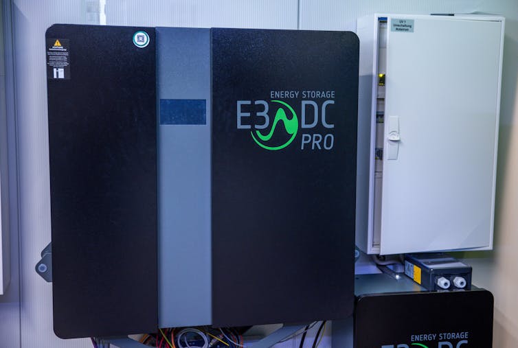
We dubbed it Merlin after King Arthur’s legendary magician to reflect the magical nature of the system: It automatically collects data on how people use energy in their homes and identifies opportunities to charge and discharge home battery storage. And it does so in a way that you always have power for whatever you need. So your air conditioning is always available, but at the same time it reduces the strain on the grid – for example, during afternoon peaks.
If demand outstrips the available generation, utilities typically ask customers to adjust their thermostats and otherwise reduce their loads. If that’s not sufficient, blackouts are possible. This is where Merlin comes in. By managing energy use in homes more intelligently, Merlin helps balance the energy supply, making electric grids more stable and reliable. Merlin manages the grid’s use of the home’s battery while maintaining the home’s normal consumption of energy.
Why it matters
To address climate change, society needs to transition to generating electrical power exclusively using nonfossil fuel sources like solar, wind and nuclear. Also, all household appliances, or end uses – heating, cooking, clothes-drying – must be electrified. A similar transition is happening with cars, moving from internal combustion engine vehicles to battery electric vehicles. However, most renewable energy sources are non-dispatchable, meaning power companies can’t just turn them on when needed.
Advertisement
This requires a fundamental shift from a centralized energy system where a power plant generates all needed electricity, to a more decentralized or distributed one. In decentralized systems, power is generated at the edges of the grid – for example, in homes with solar panels – and homes and office buildings can store energy in batteries.
Homes and office buildings also actively try to reduce or shift their loads to reduce their demands on the grid. This means fewer blackouts and less wasted energy. Plus, using energy more efficiently helps reduce greenhouse gas emissions.
What other research is being done
Researchers are working on various ways to make buildings smarter and better at shifting their energy use. In fact, the U.S. Energy Department has created a national road map to develop such grid-interactive efficient buildings to triple the energy efficiency and energy demand flexibility of buildings by 2030. In 2021, the department funded 10 projects of public-private partnerships with its Connected Communities program to develop and test technologies for grid-interactive buildings.
All these programs and developments focus on advanced control systems and promoting the adoption of smart technologies to optimize energy use, similar to Merlin.
What’s next
The next step is to test systems like Merlin in more communities under realistic conditions, and understand how well they work in different places and conditions. It is important that we gather feedback about users’ experiences and integrate them into our next prototypes to ensure the acceptability of AI systems managing people’s home energy. We are aiming to make these systems user-friendly and affordable so that everyone can benefit from smarter, greener homes.
The goal is for every neighborhood to have homes that share energy like a team, always making sure there’s enough power for everyone and using as little of it as possible from the grid.
Advertisement
The Research Brief is a short take on interesting academic work.
The Food and Drug Administration implemented a rule to go into effect on Sept. 10, 2024, requiring mammography facilities to notify women about their breast density. The goal is to ensure that women nationwide are informed about the risks of breast density, advised that other imaging tests might help find cancers and urged to talk with their doctors about next steps based on their individual situation.
The FDA originally issued the rule on March 10, 2023, but extended the implementation date to give mammography facilities additional time to adhere.
Breast density is categorized into four categories: fatty, scattered tissue, heterogeneously dense or extremely dense.
Dense breasts are composed of more fibrous, connective tissue and glandular tissue – meaning glands that produce milk and tubes that carry it to the nipple – than fatty tissue. Because fibroglandular tissue and breast masses both look white on mammographic images, greater breast density makes it more difficult to detect cancer. Nearly half of all American women are categorized as having dense breasts.
The FDA now requires specific language to ensure that all women receive the same “accurate, complete and understandable breast density information.” After a mammogram, women must be informed:
– That for those with dense breasts, additional imaging tests might help find cancer
They must also be advised to discuss their individual situation with their health care provider, to determine which, if any, additional screening might be indicated.
The FDA standardized the information women must receive. It is written at an eighth grade reading level and may address racial and literacy-level differences in women’s knowledge about breast density and reactions to written notifications.
Advertisement
For instance, our research team found disproportionately more confusion and anxiety among women of color, those with low literacy and women for whom English was not their first language. And some women with low literacy reported decreased future intentions to undergo mammographic screening.
What is the value of additional screening?
Standard mammograms use X-rays to produce two-dimensional images of the breast. A newer type of mammography imaging called tomosynthesis produces 3D images, which find more cancers among women with dense breasts. So, researchers and doctors generally agree that women with dense breasts should undergo tomosynthesis screening when available.
There is still limited scientific evidence to guide recommendations for supplemental breast screening beyond standard mammography or tomosynthesis for women with dense breast tissue. Data shows that supplemental screening with ultrasound, MRI or contrast-enhanced mammography may detect additional cancers, but there are no prospective studies confirming that such additional screening saves more lives.
So far, there is no data from randomized clinical trials showing that supplemental breast MRIs, the most often-recommended supplemental screening, reduce death from breast cancer.
Advertisement
However, more early stage – but not late-stage – cancers are found with MRIs, which may require less extensive surgery and less chemotherapy.
Various professional organizations and experts interpret the available data about supplemental screening differently, arriving at different conclusions and recommendations. An important consideration is the woman’s individual level of risk, since emerging evidence suggests that women whose personal risk of developing breast cancer is high are most likely to benefit from supplemental screening.
Because personal risk of breast cancer is a crucial consideration in deciding whether to undergo supplemental screening, women should understand their own risk.
Advertisement
The American College of Radiology recommends that all women undergo risk assessment by age 25. Women and their providers can use risk calculators such as Tyrer-Cuzick, which is free and available online.
Women should also understand that breast density is only one of several risks for breast cancer, and some of the others can be modified. Engaging in regular physical activity, maintaining a healthy weight, limiting alcohol use and eating a healthy diet rich in vegetables can all decrease breast cancer risk.
Are there potential harms?
Amid the debate about the benefits of supplemental breast screening, there is less discussion about its possible harms. Most common are false alarms: results that suggest a finding of cancer that require follow-up testing. Less commonly, a biopsy is needed, which may lead to short-term fear and anxiety, medical bills or potential complications from interventions.
MRI screening also involves use of a chemical substance called gadolinium to improve imaging. Although tiny amounts of gadolinium are retained in the body, the FDA considers the contrast agent to be safe when given to patients with normal kidney function.
MRIs may also identify incidental findings outside the breast – such as in the lungs – that warrant additional concern, testing and cost. Women should consider their tolerance for such risks, relative to their desire for the benefits of additional screening.
The out-of-pocket cost of additional screening beyond a mammogram is also a consideration; only 29 states plus the District of Columbia require insurance coverage for supplemental breast cancer screening, and only three states – New York, Connecticut and Illinois – mandate insurance coverage with no copays.
To address this knowledge deficit in some health care settings, radiologists, whose screening guidelines are more stringent than some other organizations, sometimes provide a recommendation for supplemental screening as part of their mammography report to the provider who ordered the mammogram.
Learning more about the topic in advance of a discussion with a provider can help a woman better understand her options.
Armed with information about the complexities of breast density and its impact on breast cancer screening, women can discuss their personal risk with their providers and evaluate the options for supplemental screening, with consideration of how they value the benefits and harms associated with different tests.
What happens to a dead body in an extremely cold environment? Does it decompose? How do these conditions affect how forensic scientists understand when the person died?
Estimating time of death, also called the post-mortem interval, is a complex task. It plays an important role in forensic investigations, as it can provide critical insights into the timeline of events leading up to a person’s death. This information can narrow down potential scenarios and suspects, aiding in the resolution of criminal cases.
A multitude of factors are at play at a death scene, ranging from environmental conditions to the individual’s health status prior to death. Historically, scientists have estimated time of death by observing post-mortem physical and biological changes in the body, such as stiffening, fluid collection and cooling.
These methods are limited, however, by their variability and dependence on external factors. Calculating the post-mortem interval became more precise with the advent of molecular biology. But it’s still a challenging task, especially in extreme cold weather conditions. There is often a lack of obvious signs of decomposition on a frozen body during the first months after death.
Advertisement
We are forensic scientists leading the forensics programs at the University of North Dakota and the University of Central Lancashire. We use molecular biology and bioinformatics to develop tools to help researchers and investigators more accurately estimate the post-mortem interval. Our recently published research in Frontiers in Microbiology found that studying the microbes involved in decomposition could predict time elapsed since death in extreme cold conditions with high accuracy.
In an extremely cold environment like North Dakota’s winters, traditional methods might not be enough to understand decomposition and estimate time of death. For instance, the body cools much faster in cold conditions, which can skew estimates based on body temperature.
Similarly, cold environments can delay the onset and duration of rigor mortis, or body stiffening. The process of decomposition, including the activity of insects and other scavengers that contribute to the breakdown of the body, can also be slowed or halted by freezing temperatures.
Snow is another important factor when investigating decomposition. It can insulate a body by trapping residual heat and raising its temperature slightly higher than the surrounding environment. This insulating effect allows the body to decompose at a slower rate compared with bodies exposed to open air.
Microbes and time since death
In conditions of extreme cold, it becomes necessary to employ additional means to understand decomposition and estimate the time of death. Advanced molecular techniques, such as analyzing the microbiome, gene expression and protein degradation, can help provide valuable information about the crime scene.
Each organism has distinct microbial characteristics that act like a fingerprint. The necrobiome, a community of microbes associated with decomposing remains, plays a crucial role in decay. Specific microbes are present during different stages of decomposition, contributing to the breakdown of tissues and the recycling of nutrients. Forensic investigators can sample what microbes are living in a dead body to deduce how long ago a person died based on the makeup of the microbial population.
Advertisement
Our study focused on identifying common patterns in the microbial changes that occur during decomposition in extreme cold environments. Over a period of 23 weeks, we collected and analyzed 393 samples of microbes from the inside and outside of the noses dead pigs covered in snow. Pigs decompose similarly to humans and are commonly used in forensic research. We developed models to estimate the post-mortem interval by pairing microbial genetic data with environmental data such as snow depth and outdoor temperature.
Overall, we found that the bacterial species Psychrobacter, Pseudomonas and Carnobacterium may best predict time after death in extreme winter conditions up to six months after death, with a margin of error of just over nine days.
We found that different bacterial species are most abundant at different time intervals. For example, levels of Psychrobacter increase five weeks after death and are most abundant at 10 weeks, while Pseudomonas increase between five to nine weeks and hit a peak at 18 weeks.
Advertisement
Improving forensics
Death is often an unpleasant topic to bring into a conversation. But from a forensic perspective, having techniques and methods to determine when someone has died can help bring justice and peace for loved ones.
Our study found that decomposition does not completely halt even in cold environments. Studying the microenvironment – the local conditions surrounding the body, including temperature, humidity and microbial activity – can reveal crucial information about the decomposition process. The key microbial species we identified served as biomarkers of death, allowing us to develop time-of-death models that researchers can use to overcome the limitations of just visually examining remains.
Microbes can become a crucial piece of the puzzle during the process of investigating a death by aiding in constructing more precise timelines, even in extreme conditions.
The Food and Drug Administration implemented a rule to go into effect on Sept. 10, 2024, requiring mammography facilities to notify women about their breast density. The goal is to ensure that women nationwide are informed about the risks of breast density, advised that other imaging tests might help find cancers and urged to talk with their doctors about next steps based on their individual situation.
The FDA originally issued the rule on March 10, 2023, but extended the implementation date to give mammography facilities additional time to adhere.
Breast density is categorized into four categories: fatty, scattered tissue, heterogeneously dense or extremely dense.
Advertisement
Dense breasts are composed of more fibrous, connective tissue and glandular tissue – meaning glands that produce milk and tubes that carry it to the nipple – than fatty tissue. Because fibroglandular tissue and breast masses both look white on mammographic images, greater breast density makes it more difficult to detect cancer. Nearly half of all American women are categorized as having dense breasts.
The FDA now requires specific language to ensure that all women receive the same “accurate, complete and understandable breast density information.” After a mammogram, women must be informed:
– That for those with dense breasts, additional imaging tests might help find cancer
Advertisement
They must also be advised to discuss their individual situation with their health care provider, to determine which, if any, additional screening might be indicated.
The FDA standardized the information women must receive. It is written at an eighth grade reading level and may address racial and literacy-level differences in women’s knowledge about breast density and reactions to written notifications.
Advertisement
For instance, our research team found disproportionately more confusion and anxiety among women of color, those with low literacy and women for whom English was not their first language. And some women with low literacy reported decreased future intentions to undergo mammographic screening.
What is the value of additional screening?
Standard mammograms use X-rays to produce two-dimensional images of the breast. A newer type of mammography imaging called tomosynthesis produces 3D images, which find more cancers among women with dense breasts. So, researchers and doctors generally agree that women with dense breasts should undergo tomosynthesis screening when available.
There is still limited scientific evidence to guide recommendations for supplemental breast screening beyond standard mammography or tomosynthesis for women with dense breast tissue. Data shows that supplemental screening with ultrasound, MRI or contrast-enhanced mammography may detect additional cancers, but there are no prospective studies confirming that such additional screening saves more lives.
So far, there is no data from randomized clinical trials showing that supplemental breast MRIs, the most often-recommended supplemental screening, reduce death from breast cancer.
Advertisement
However, more early stage – but not late-stage – cancers are found with MRIs, which may require less extensive surgery and less chemotherapy.
Various professional organizations and experts interpret the available data about supplemental screening differently, arriving at different conclusions and recommendations. An important consideration is the woman’s individual level of risk, since emerging evidence suggests that women whose personal risk of developing breast cancer is high are most likely to benefit from supplemental screening.
Because personal risk of breast cancer is a crucial consideration in deciding whether to undergo supplemental screening, women should understand their own risk.
Advertisement
The American College of Radiology recommends that all women undergo risk assessment by age 25. Women and their providers can use risk calculators such as Tyrer-Cuzick, which is free and available online.
Women should also understand that breast density is only one of several risks for breast cancer, and some of the others can be modified. Engaging in regular physical activity, maintaining a healthy weight, limiting alcohol use and eating a healthy diet rich in vegetables can all decrease breast cancer risk.
Are there potential harms?
Amid the debate about the benefits of supplemental breast screening, there is less discussion about its possible harms. Most common are false alarms: results that suggest a finding of cancer that require follow-up testing. Less commonly, a biopsy is needed, which may lead to short-term fear and anxiety, medical bills or potential complications from interventions.
MRI screening also involves use of a chemical substance called gadolinium to improve imaging. Although tiny amounts of gadolinium are retained in the body, the FDA considers the contrast agent to be safe when given to patients with normal kidney function.
MRIs may also identify incidental findings outside the breast – such as in the lungs – that warrant additional concern, testing and cost. Women should consider their tolerance for such risks, relative to their desire for the benefits of additional screening.
The out-of-pocket cost of additional screening beyond a mammogram is also a consideration; only 29 states plus the District of Columbia require insurance coverage for supplemental breast cancer screening, and only three states – New York, Connecticut and Illinois – mandate insurance coverage with no copays.
To address this knowledge deficit in some health care settings, radiologists, whose screening guidelines are more stringent than some other organizations, sometimes provide a recommendation for supplemental screening as part of their mammography report to the provider who ordered the mammogram.
Learning more about the topic in advance of a discussion with a provider can help a woman better understand her options.
Armed with information about the complexities of breast density and its impact on breast cancer screening, women can discuss their personal risk with their providers and evaluate the options for supplemental screening, with consideration of how they value the benefits and harms associated with different tests.